































Table of Contents
- Key Points
- Understanding Image Segmentation and Graph Theory
- Solving Image Segmentation Problems with Multi-Cut
- How to Implement the Multi-Cut Segmentation
- Pros and Cons of the Multi-Cut Approach
- Frequently Asked Questions
- Further Explorations



Understanding Image Segmentation and Graph Theory
The Importance of Image Segmentation
Image segmentation is the process of partitioning a digital image into multiple segments (sets of pixels). More precisely, Image Segmentation is the process of assigning a label to every pixel in an image such that pixels with the same label share certain visual characteristics.
The result of image segmentation is a set of segments that collectively cover the entire image. Each of the pixels in a region are similar with respect to some characteristic or computed property, such as color, intensity, or texture. It is a vital task in numerous applications, such as autonomous driving, medical imaging, and object recognition. In autonomous driving, image segmentation allows vehicles to identify and classify different parts of the road scene, such as road surfaces, cars, pedestrians, and traffic signs. In medical imaging, it enables the analysis of structures such as tumors from imaging data which may come from CT scans, MRI scans and X-rays. Accurately segmenting images is crucial for effective analysis and decision-making in these and many other fields.
The goal of image segmentation is to simplify and/or change the representation of an image into something that is more Meaningful and easier to analyze.
Image segmentation is used to locate objects and boundaries (lines, curves, etc.) in images. More precisely, image segmentation is the process of assigning a label to every pixel in an image, such that pixels with the same label share certain characteristics. The result of image segmentation is a set of segments that collectively cover the entire image, or a set of contours extracted from the image (see edge detection). Each of the pixels in a region are similar with respect to some characteristic or computed property, such as color, intensity, or texture. Adjacent regions are significantly different with respect to the same characteristic(s). When applied to a stack of images, typical in MRI/CT scanning, the resulting contours after image segmentation can be used to create 3D reconstructions with the help of interpolation algorithms like marching cubes.
Key Concepts for Image Segmentation:
- Pixels and Regions: Understanding the fundamental units of an image, pixels, and how they form larger, meaningful regions.
- Image Properties: Identifying and utilizing image characteristics like color, intensity, and texture to distinguish between different segments.
- Applications: Recognizing the diverse real-world applications of image segmentation, from self-driving cars to medical diagnoses.
How Graph Theory Comes into Play
Graph theory provides a powerful framework for tackling image segmentation problems. At its core, graph theory deals with the study of graphs, which are mathematical structures used to model pairwise relations between objects. A graph consists of vertices (nodes) and edges that connect these vertices.
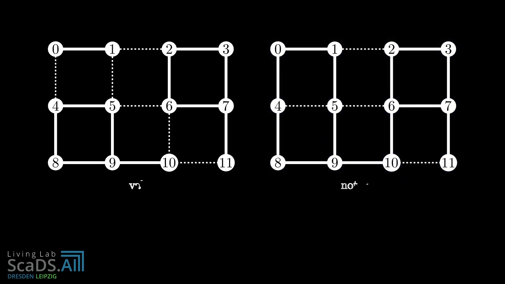
In image segmentation, we can represent an image as a graph where each pixel corresponds to a vertex, and edges connect neighboring pixels. The weight of each edge represents the similarity between the connected pixels; the more similar two pixels are (in terms of intensity, color, or texture) the higher the edge weight. This transforms the image segmentation task into a graph partitioning problem.
Building the Image Graph:
- Pixel as Vertices: Each pixel in the image becomes a vertex in the graph.
- Connectivity: Neighboring pixels are connected by edges (4-connectivity or 8-connectivity are common).
- Edge Weights: Edges are assigned weights based on the similarity between connected pixels. High similarity results in high weight.
Graph Multi-Cut Problem then are used to solve the image segmentation problem.
What is Graph Multi-Cut Problem
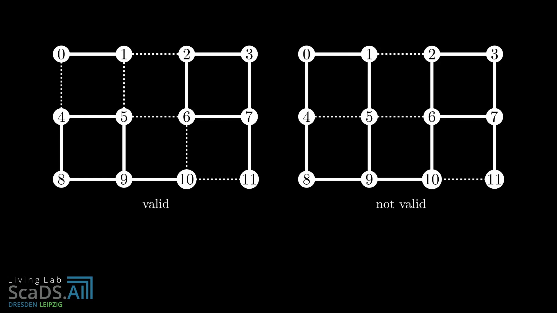
In graph theory, a multi-cut is a set of edges whose removal disconnects a graph into multiple connected components. The graph multi-cut problem aims to find a multi-cut that minimizes the total weight of the removed edges while ensuring that specific pairs of vertices (terminal pairs) are separated into different components. Think of it as strategically cutting an image graph to isolate different objects or regions.
Mathematical Definition:
Given a graph G = (V, E) with edge weights and a set of terminal pairs, the multi-cut problem involves finding a subset of edges that minimizes the sum of their weights, such that each terminal pair is disconnected in the resulting graph.
Connection to Image Segmentation:
By formulating image segmentation as a graph multi-cut problem, we aim to find the optimal way to "cut" the image graph to isolate different objects based on their dissimilarities, while keeping similar pixels within the same segment.
Solving Image Segmentation Problems with Multi-Cut
Formulating the Multi-Cut Problem for Neural Tissue Segmentation
To segment electron microscopy images of neural tissue using a multi-cut approach, we need to define:
-
Vertices: Each pixel or small group of pixels in the electron microscopy image becomes a vertex in the graph.
-
Edges: Connect neighboring vertices.
-
Edge Weights: This is where deep learning steps in. A pre-trained deep neural network predicts the likelihood of a boundary (membrane) between neighboring pixels. This prediction is used to calculate the weights of the edges. Higher probability of a membrane means a lower (more negative) weight, indicating that the edge is a good candidate for cutting.
-
Terminal Pairs: Not always explicitly defined. Can use heuristics based on neural net output.
This process transforms the image into a format suitable for processing.
Solving Multi-Cut Problems: Algorithms
Solving the multi-cut problem is NP-hard, meaning that finding the optimal solution can be computationally expensive for large graphs. Therefore, various algorithms are employed to find near-optimal solutions within a reasonable time.
-
Greedy Algorithms: A straightforward approach is to iteratively remove edges with the lowest weights (highest membrane likelihood) until the terminal pairs are disconnected. This is fast but may not always produce the best results.
-
Integer Linear Programming (ILP): The multi-cut problem can be formulated as an ILP problem, which can then be solved using ILP solvers. This method guarantees an optimal solution but is computationally intensive.
-
Specialized Algorithms: Advanced algorithms, often based on message-passing or other approximation techniques, are designed to handle large graphs efficiently. One method named Mutex Watershed Algorithm, as seen in the YouTube video.
These all produce fast and meaningful results.
The Role of Deep Neural Networks
Deep neural networks significantly enhance image segmentation by providing accurate boundary predictions. Instead of relying on simple image features, deep learning models can learn complex Patterns and contextual information from the data. For electron microscopy images, a convolutional neural network (CNN) can be trained to identify the presence of membranes between cells. This is done by providing training data containing image patches and corresponding labels indicating whether a membrane exists at the center pixel.
The trained CNN can then predict the likelihood of a membrane at each pixel in a new image.
- Feature Extraction: Deep learning models learn Relevant features from the image data.
- Boundary Prediction: The neural network predicts the probability of a membrane between pixels.
- Edge Weight Calculation: These probabilities are converted into edge weights for the graph multi-cut problem.








Most people like

































Report
Please select a reason for reporting
- 13 Reasons to Switch from Snapchat to Text Messaging
- 7 Powerful Speech to Text Apps to Boost Productivity
- 10 Best Speech to Text Tools for Effortless Transcription
- 13 Free Tools to Easily Transcribe Audio to Text
- 15 Surprising Ways Google's Speech-to-Text Boosts Productivity
- 7 Best Free Online Audio to Text Transcription Tools
- 9 Tips to Transcribe Speech to Text Faster and Better
- 13 Best Speech to Text Software for Windows 10 in 2023
- 7 Tips for Choosing the Best Transcriber for Audio to Text
- 11 Reasons Why Dragon Speech-to-Text Apps are Game-Changers
- 6 Secrets to Enhance Your Speech to Text Transcription
- 6 Brilliant Ways to Boost Productivity with Talk-to-Text on Your MacBook
- 6 Ways Transcription Software Converts Audio to Text
- 8 Tips to Transcribe Speech to Text Like a Pro
- 6 Reasons IBM Watson Speech to Text is a Game-Changer
- 14 Powerful Ways Talk-to-Text Improves Your Workflow
- 12 Top Windows Speech-to-Text Software for Efficient Transcription
- 12 Game-Changing Benefits of Apple's Speech to Text Technology
- 9 Powerful Use Cases for AWS Speech to Text Technology
- 10 Easy Steps to Transcribe YouTube Videos to Text
- transcription audio en texte
- online audio transcription
- free audio file transcription
- free ai audio transcription
- audio transcription service
- audio transcription online free
- audio transcription free online
- audio to transcription software
- audio and transcript
- ai audio transcription free
- voice recording to text converter
- translate voice recording to text
- record voice to text
- audio recording to text converter
- voice recording and transcription
- chatgpt voice to text
- gmail voice to text
- speech to text reader
- speech to text ai free
- speech to talk
- How SEO and Marketing Dashboards Empower Agencies to Drive Results
- How to Use AI Tools to Find Internal Linking Opportunities Across a Business Website
- Discover Your Perfect Haircut with AI Hairstyle Apps
- Free Reputation Calculator Tools: How They're Adapting to Score AI Visibility, Not Just Rankings
- Design Professional Flyers for Free on Your Phone
- Best Free Flyer Maker Apps for Android: Design Like a Pro
- How to Know If Your Business Is Ready for an AI Customer Service Agent
- Revolutionizing Storytelling: From Novels to Screenplays with AI
- Create Engaging Animated Explainer Videos with a Free AI Tool
- Stop Multi-Subscribing: Benchmark Top AI Visual Models in One Canvas
- ASOMobile Helps Teams Benchmark App Store Screenshots Against Competitors
- How to Match LMS Software With Your Organization's Training Plan
- Revolutionizing Email Marketing Agency with AI
- How to Convert BTC to HNT
- Unleashing the Power of AI: Cognizant's Roadmap for Transformation
- Frame to Video Review: A Practical Look at Turning Two Still Frames Into Motion
- NVIDIA NIM: Revolutionizing Supply Chain Data Analysis with AI
- Top AI Tools That Help Brands Stand Out Offline (Not Just Online)
- Top 5 Cold Email Software: Maximize Your Outreach in 2025
- Magic School AI: Revolutionizing Quiz & Assessment Generation

Copyright ©2026 toolify