






Table of Contents
- Key Points
- Understanding Deepmedic Architecture for Liver Segmentation
- How to Use Deepmedic
- Pricing for Deepmedic Software
- Advantages and Disadvantages of Using Deepmedic for Liver Segmentation
- Core Features of Deepmedic
- Key Use Cases for Deepmedic
- Frequently Asked Questions about Deepmedic
- Related Questions About Medical Segmentations



Understanding Deepmedic Architecture for Liver Segmentation
An In-depth Look at the Three-Channel CNN
Deepmedic distinguishes itself by employing a three-channel convolutional neural network (CNN) architecture specifically tailored for liver segmentation in CT images. This multi-channel approach allows the network to capture information at varying resolutions, enabling a more comprehensive understanding of the liver's anatomy and its surrounding context.
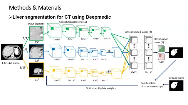
The core idea is to combine the strengths of high-resolution and low-resolution pathways to achieve a balance between detailed feature extraction and contextual awareness.
- High-Resolution Channel: This channel processes the input image at its original resolution. It's designed to capture fine-grained details within the liver, such as subtle variations in tissue density or the presence of small lesions. This is crucial for accurate boundary delineation and identifying intricate structural features. The CNN architecture will extract high level features after running the patches sampled from the input.
- Downsampled Channels: To provide contextual information, the architecture includes two additional channels that process downsampled versions of the input image. These channels capture the broader anatomical context surrounding the liver, allowing the network to understand the liver's relationship to neighboring organs and structures. This contextual awareness helps to reduce false positives and improve the overall robustness of the segmentation. The input image is downsampled by factors of 1/5 and 1/15.
By fusing information from these three channels, Deepmedic can effectively leverage both local details and global context for precise liver segmentation. This design is instrumental in handling the complexities of liver anatomy and the variability present in CT images. The convolutional layers are used in 10 layers.
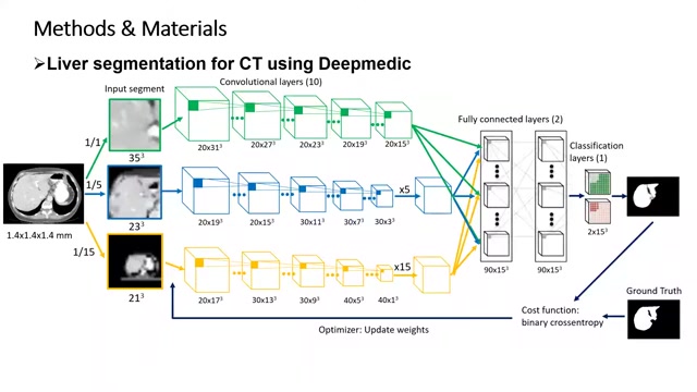
Fully Connected Layers Fully connected layers (2 layers) are utilized to combine features together. The high and lower resolution features are combined.
Classification Layers Classification layers (1 layer) are used to output a probability map. The output represents the probability of each voxel belonging to the liver.
Binary Cross Entropy Cost Function The similarity between the ground truth and probability map is combined using the binary cross entropy cost function.
Optimizer An optimizer is used to update the weights in the network so that the cost function can be minimized.
Input Segment and Patch Extraction
The process begins with an input segment, which represents a portion of the CT scan encompassing the liver region. To efficiently process this segment, Deepmedic extracts small patches, or sub-volumes, from the input image. These patches serve as the fundamental units of analysis for the CNN.
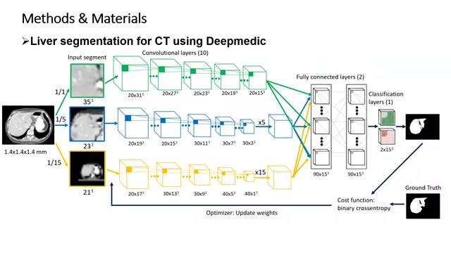
These image patches are extracted from these downsampled images. The high-resolution channel utilizes input segments from the images with original resolution. It then extracts high-level features from these segments. The second and third channels use patches downsampled from the images.
The extraction of smaller patches allows the network to focus on local features while reducing the computational burden. The size of these patches is carefully chosen to balance the need for sufficient contextual information with the limitations of available memory and processing power. Patch extraction helps in extracting high-level and low-level features.
The patches are then fed into the three-channel CNN, where they undergo a series of convolutional, pooling, and non-linear activation operations. These operations extract relevant features from the patches, gradually building a hierarchical representation of the liver anatomy.
Downsampling is done to provide contextual information to the model. These different input types are combined in the fully connected layers.
Convolutional Layers and Feature Extraction
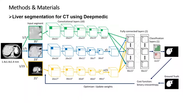
Convolutional layers form the heart of Deepmedic's feature extraction process. These layers apply a set of learnable filters to the input patches, convolving them to produce feature maps that highlight specific characteristics within the image. Each convolutional layer learns to detect different features, such as edges, textures, and patterns. The stacking of multiple convolutional layers allows the network to learn increasingly complex and abstract features, gradually building a hierarchical representation of the liver anatomy.
After each convolutional layer, a non-linear activation function is applied to introduce non-linearity into the network. This non-linearity is essential for learning complex relationships between features and enabling the network to model the intricate variability present in medical images. Common activation functions include ReLU (Rectified Linear Unit) and its variants.
High Level Feature Extractions: High level features are extracted using a series of convolutional layers in order to extract features.
Key Use Cases for Deepmedic
Applications in Clinical Settings
Deepmedic's liver segmentation capabilities find application in various clinical scenarios:
- Surgical Planning: Assisting surgeons in pre-operative planning by providing accurate anatomical information.
- Disease Diagnosis: Aiding in the diagnosis of liver diseases by facilitating the detection and quantification of abnormalities.
- Treatment Monitoring: Tracking the effectiveness of treatment by monitoring changes in liver volume and structure.
- Research: Serving as a valuable tool for researchers studying liver anatomy, pathology, and treatment outcomes.
It can significantly help with liver analysis and surgical planning through its accurate segmentation.



Most people like


































Report
Please select a reason for reporting
- 13 Reasons to Switch from Snapchat to Text Messaging
- 7 Powerful Speech to Text Apps to Boost Productivity
- 10 Best Speech to Text Tools for Effortless Transcription
- 13 Free Tools to Easily Transcribe Audio to Text
- 15 Surprising Ways Google's Speech-to-Text Boosts Productivity
- 7 Best Free Online Audio to Text Transcription Tools
- 9 Tips to Transcribe Speech to Text Faster and Better
- 13 Best Speech to Text Software for Windows 10 in 2023
- 7 Tips for Choosing the Best Transcriber for Audio to Text
- 11 Reasons Why Dragon Speech-to-Text Apps are Game-Changers
- 6 Secrets to Enhance Your Speech to Text Transcription
- 6 Brilliant Ways to Boost Productivity with Talk-to-Text on Your MacBook
- 6 Ways Transcription Software Converts Audio to Text
- 8 Tips to Transcribe Speech to Text Like a Pro
- 6 Reasons IBM Watson Speech to Text is a Game-Changer
- 14 Powerful Ways Talk-to-Text Improves Your Workflow
- 12 Top Windows Speech-to-Text Software for Efficient Transcription
- 12 Game-Changing Benefits of Apple's Speech to Text Technology
- 9 Powerful Use Cases for AWS Speech to Text Technology
- 10 Easy Steps to Transcribe YouTube Videos to Text
- transcription audio en texte
- online audio transcription
- free audio file transcription
- free ai audio transcription
- audio transcription service
- audio transcription online free
- audio transcription free online
- audio to transcription software
- audio and transcript
- ai audio transcription free
- voice recording to text converter
- translate voice recording to text
- record voice to text
- audio recording to text converter
- voice recording and transcription
- chatgpt voice to text
- gmail voice to text
- speech to text reader
- speech to text ai free
- speech to talk
- Meshy AI: How to Create 3D Models from Text and Images
- How SEO and Marketing Dashboards Empower Agencies to Drive Results
- How to Use AI Tools to Find Internal Linking Opportunities Across a Business Website
- Discover Your Perfect Haircut with AI Hairstyle Apps
- Free Reputation Calculator Tools: How They're Adapting to Score AI Visibility, Not Just Rankings
- Design Professional Flyers for Free on Your Phone
- Best Free Flyer Maker Apps for Android: Design Like a Pro
- How to Know If Your Business Is Ready for an AI Customer Service Agent
- Revolutionizing Storytelling: From Novels to Screenplays with AI
- Create Engaging Animated Explainer Videos with a Free AI Tool
- Automate Lead Generation: A Comprehensive Guide with AI Agents
- Revideo.io: The Future of Reverse Video Search Technology
- Revolutionizing Email Marketing Agency with AI
- Breaking the Character Drift Dilemma: A Practical Test of Smart Shot
- Meshy AI: How to Create 3D Models from Text and Images
- ASOMobile Helps Teams Benchmark App Store Screenshots Against Competitors
- How to Match LMS Software With Your Organization's Training Plan
- How to Convert BTC to HNT
- Unleashing the Power of AI: Cognizant's Roadmap for Transformation
- Frame to Video Review: A Practical Look at Turning Two Still Frames Into Motion

Copyright ©2026 toolify